To study nucleic acids and proteins, including DNA and RNA, you will more than likely use what is referred to as gel electrophoresis. It is a commonly used application when it comes to food sciences, life sciences, and even mining.
Many people may be wondering how does a gel electrophoresis chamber work? To understand this, we should begin with learning about how a gel electrophoresis chamber works.
How Does a Gel Electrophoresis Chamber Work?
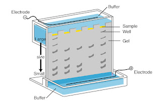
When separating nucleic acids or proteins using the gel electrophoresis method, you have to run the samples through a gel electrophoresis chamber. It features a cathode on one end and an antinode on the other in a platform that will hold the gel mixture in the center. A buffer is then added that will create a charge gradient when an electric current is applied since the buffer will keep the gel cool and stop it from getting too hot.
The molecules will then gravitate towards the positive electrode when the electric current is applied, and the smaller molecules will move through the gel matrix faster than the bigger ones. After this process is completed, you will be able to see bands of proteins and nucleic acids that have been separated based on their molecular weights.
What is Horizontal Electrophoresis?
When it comes to horizontal electrophoresis systems, which are also called submarine units, they are systems that have been created to run both agarose and polyacrylamide gels that have been submerged in a running buffer. The samples are then put into an electronic field so they can migrate to the anode or cathode depending on how they are charged. These systems can be utilized to separate proteins as well as DNA and RNA in a laboratory so that they can be screened quickly for sample quantification, PCR amplification detection, and for determining size.
These systems normally come with a submarine tank along with Combs, electrodes, a power supply, and a casting tray. There are several sizes available that can handle small, medium, and large-sized gels. The size choice will be determined by the sample type and size as well as the running time needed for the sample in the electrophoresis conditions. Pulse fields can also be used for genotyping, fingerprinting, and epidemiological studies for a variety of pathogenic organisms. You should consider what type of sample you are running along with the time and voltage needed to separate the fragments, the benchtop size restrictions that are in place, and the conditions of the buffer.
What is Vertical Electrophoresis?
Vertical gel electrophoresis is more complex than the horizontal method when it comes to its set-up. This type of system is used to separate proteins instead of nucleic acids, but before you can separate proteins, you have to cause a disruption of their quaternary structure to create a linear strand. You can achieve this by treating the protein with sodium dodecyl sulfate which will break the disulfide bond to get to the denatured proteins.
Once the proteins are in linear strands, you can then utilize the vertical gel to separate out all of the strands. The vertical gel electrophoresis method uses what is called polyacrylamide gel Because it has smaller pores. This is due to the fact that linear protein strands are smaller than those of DNA and RNA.
Key Difference Between Horizontal and Vertical Electrophoresis
Basically, gel electrophoresis is a technique used in a lab that used to separate mixtures containing DNA, RNA, and other proteins going by their different charges and molecular sizes. The method is used to separate out DNA, RNA, or proteins by running them through a gel that contains small pores.
When it comes to this process, there are two main kinds of gel electrophoresis methods, including horizontal gel electrophoresis and vertical gel electrophoresis. Both of these methods follow basically the same theory of gel electrophoresis, yet there are some differences between the two of them.
One of the main differences between the two different systems is their orientation as well as their buffer system. When it comes to horizontal gel electrophoresis, the gel matrix is submerged in a buffer that is constantly running but when using vertical gel electrophoresis, the gel is put in vertically in the buffer system does not run continuously. Also, the vertical system boasts two chambers while the vertical method only uses one chamber. This means that when you use the vertical electrophoresis system, the molecules will separate better and there will be a greater resolution.
In horizontal gel electrophoresis, an agarose gel is used while the vertical process uses acrylamide gel. The agarose gels have bigger pores than the acrylamide gels. When you want to separate mixtures that contain both are in a and DNA molecules, you should use the horizontal gel electrophoresis method but if you simply want to separate proteins from each other, you will use the vertical gel electrophoresis method.
What is the Best Method to Use?
Both methods work well for the testing processes they are designed to conduct. Vertical gel electrophoresis seems to be the most popular method for separating protein since it is easier to prepare polyacrylamide gel vertically than horizontally. However, it is normally only used on proteins, so the horizontal gel electrophoresis is actually the best method to use when you want to separate and examine strands of DNA and RNA in the testing process.